WORLD NEWS
A metal-free molecule glowed red and enhanced MRI contrast
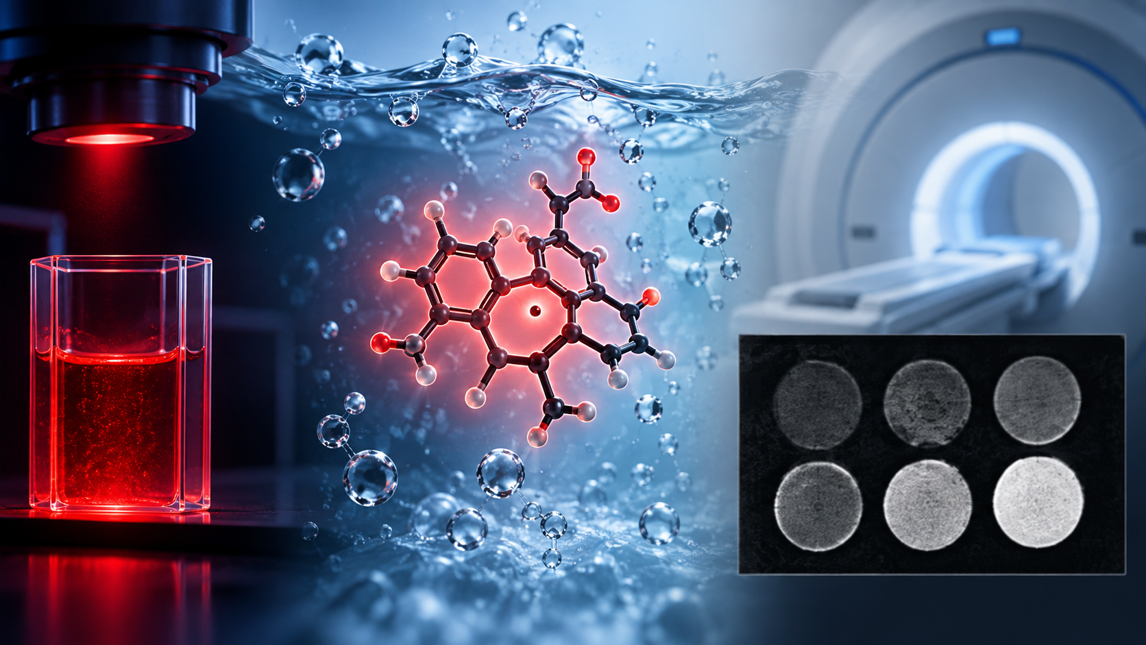
TTM-trisTP3B and TTM-trisTP9B emitted near 690 and 685 nm in water; the first remained fluorescent around living-cell nuclei for at least 24 hours.
They improved T1 contrast in MRI phantoms about 2.5–4.5 times over earlier water-soluble TTM radicals. That comparison is not a clinical image-improvement rate.
How one molecule serves two modes
Fluorescence uses absorbed and re-emitted light, while MRI reads changes in water-proton spin relaxation. Unpaired electrons in the radicals provide both luminescent and paramagnetic functions.
Why water solubility matters
Earlier emitting TTM radicals were hydrophobic. Hydrophilic groups and adjusted donor positions preserved light emission and spin behaviour in water.
Why this is not yet a gadolinium replacement
Removing metal opens a design route, but toxicity, distribution, clearance, dose, tissue contrast and long-term stability remain untested in animals and humans.
What must be tested next
Animal imaging, pharmacokinetics, organ and immune toxicity, and performance at clinical magnetic fields must establish whether the dual probe is useful.
Primary source and independent checks
Primary source: Advanced Science paper
Institutional measurement or explanation: Kyushu University research release
Independent check: PubMed bibliographic and abstract check
Read in other languages
한국어 · English · Español · 日本語 · Tiếng Việt