WORLD NEWS
They took X-rays in space after just four hours of training
Nonmedical spaceflight crewmembers acquired the first human radiographs in orbit after only four hours of training.
Images from the Fram2 flight reached diagnostic quality, but no injured patient was diagnosed and the study involved only a few participants.
What they imaged in orbit
Fram2 launched on March 31, 2025 and spent 3 days 14 hours in a 425–450 kilometre polar orbit. The crew used an ultraportable wireless digital X-ray generator and detector.
They imaged a hand, forearm, chest, abdomen and pelvis, plus a smartwatch and calibration object. Digital images were available immediately onboard.
How far four hours of training went
Three crewmembers trained and joined the study; two performed in-flight imaging because orbital time was limited. Three independent radiologists evaluated the images.
There were seven preflight and seven in-flight anatomic radiographs. Every in-flight image scored at least 3 on a five-point scale, the threshold for diagnostic quality.
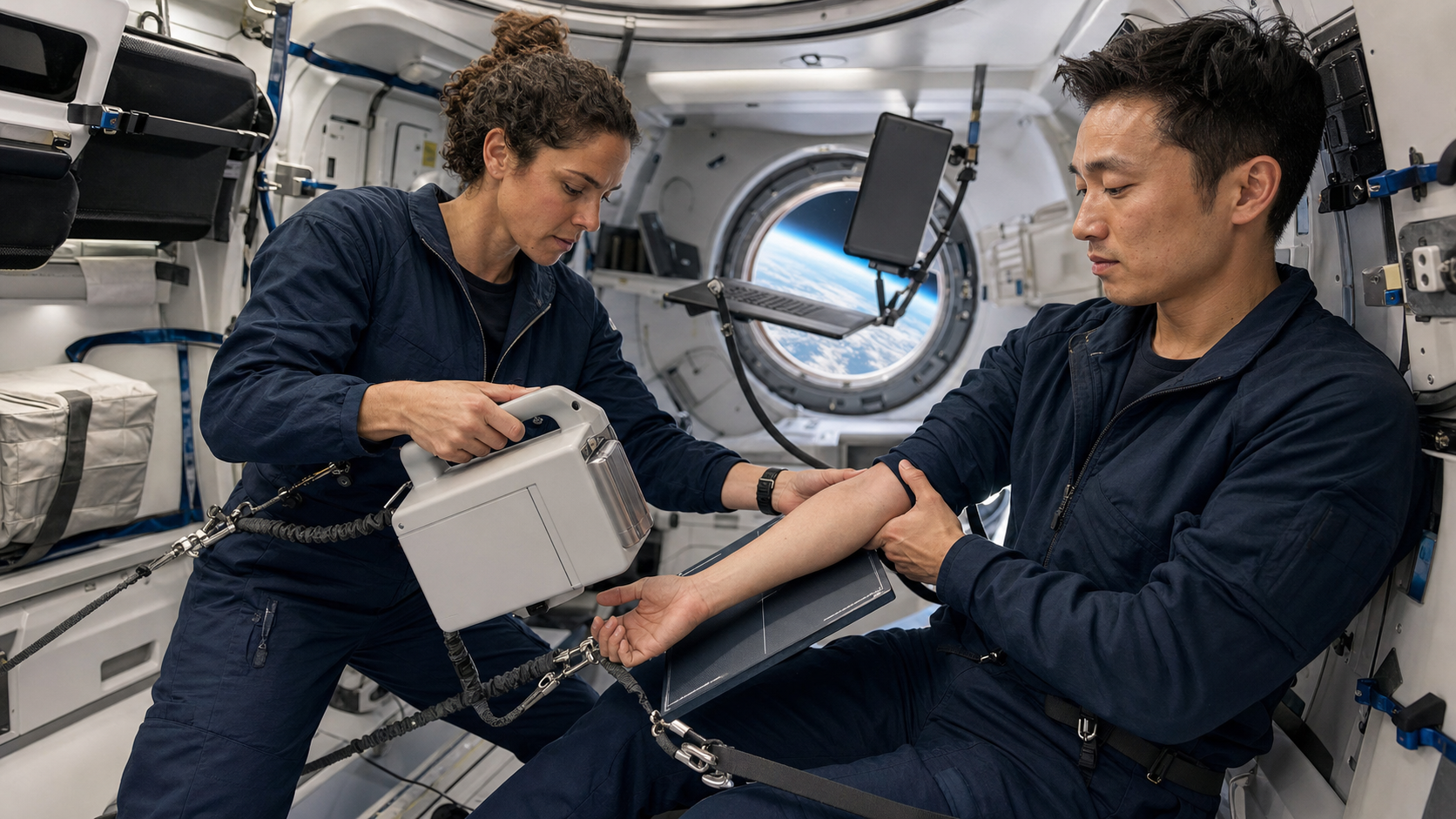
Image quality survived microgravity
Researchers found no evidence of differences in overall quality, spatial resolution or contrast resolution between preflight and in-flight images. Internal equipment details below a millimetre were visible.
Positioning of chest, abdomen and pelvis images was worse in flight. Keeping the person, source and detector aligned remains the main operational problem.
Why this matters for the Moon and Mars
For more than four decades, ultrasound was the only reliable medical imaging modality in orbit. Radiography could add information for fractures, chest problems and foreign objects.
The same system could inspect spacesuits, electronics and satellites without dismantling them.
This is not a space hospital yet
Only three people participated, and the study did not test diagnostic accuracy in real illness or injury. The device also sustained superficial damage during return.
Future flights must validate mounting, radiation management, examination rules and remote interpretation with larger samples.
Official primary sources
Primary source: Radiology original research
Primary source: Radiological Society of North America research release