WORLD NEWS
Researchers watched cellular activity from brain to kidney across a living zebrafish
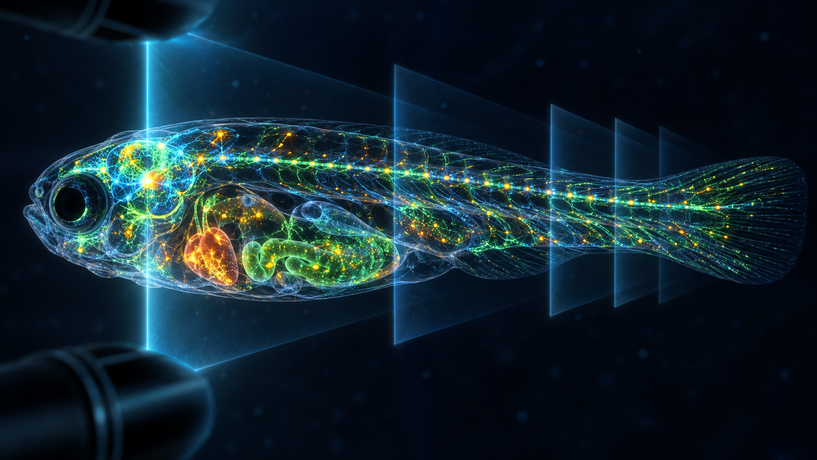
WHOLISTIC simultaneously recorded cellular activity across most of a living larval zebrafish, including brain, heart, liver, gut, pancreas, kidney and circulation. It narrows the gap between detailed single-organ and coarse whole-body imaging.
The platform was validated in small, transparent, genetically fluorescent fish. It is not ready for clinical human imaging.
How it works
Genetically encoded calcium sensors glow with activity; fast volumetric fluorescence microscopy and machine learning organize space and time.
What appeared
The team saw waves along kidney nephrons, body-wide changes during sleep-like states, ketamine-responsive cells near the brain and brainstem control of blood redistribution.
Testing cause
Optogenetic stimulation helped test whether brain signals caused body responses rather than merely occurring together.
Limits
Most of the body is not every cell. Labelling, resolution, light exposure and interpretation constrain results, and fish findings do not transfer directly to humans.
Next
Disease, immune and drug models must show which cross-organ circuits repeat and how live activity maps onto anatomy.
Primary source and independent checks
Read in other languages
한국어 · English · Español · 日本語 · Tiếng Việt