WORLD NEWS
Changes that appeared to be temperature signals of nanodiamonds in cells could be due to electric fields.
Research has drawn attention again that some of the energy transfer read as temperature changes by nanodiamond quantum sensors inside cells can also occur from changes in surrounding charges. The researchers compared surface-coated and uncoated particles to isolate the electric field signals and detect changes when macrophages respond to inflammatory stimuli.
This does not mean that the temperature inside cells does not change at all or that a new cancer diagnosis method has been completed. This is a physical experiment using cultured macrophage-like cells and a small number of particles, and the ability to accurately measure temperature and charge simultaneously in actual tissues and patients has not yet been verified.
What does nanodiamond read?
Nitrogen-vacancy defects in diamond crystals have energy levels that respond to microwaves and light. When the surrounding temperature or electric field changes, the spacing between levels also moves. The advantage is that if very small particles are placed inside a cell, changes in the cell’s environment can be tracked using light.
Why were temperature and charge confused?
Previously, the shift in zero magnetic field splitting values was mainly interpreted as being due to temperature. The new analysis showed that the transverse electric dipole effect, which occurs when charges drop or gather around a particle, can also move the same amount. So we need to revisit our interpretation of unrealistically large cell temperature changes of about 2 to 18 degrees Celsius.
What changed the coating?
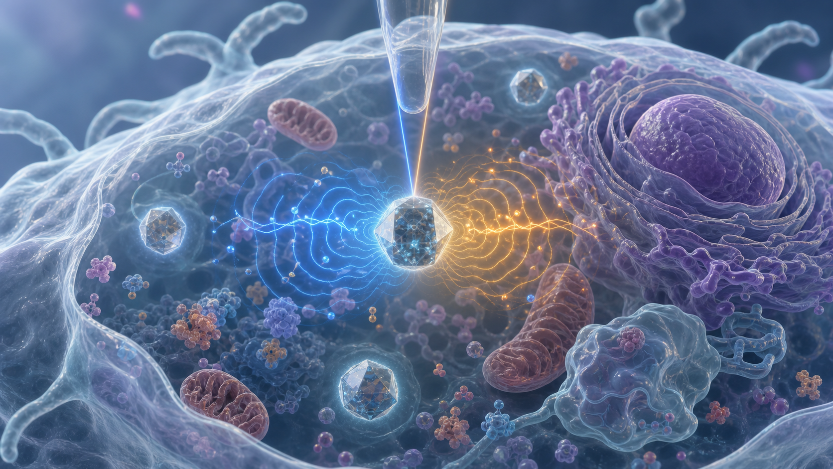
Core-shell particles coated with silica shells had smaller signal wobbles than uncoated particles. The surface coating reduces the influence of environmental charges, providing a clue to splitting the effects of temperature and electric fields. The coated particles also reduced particle-induced toxicity and inflammatory responses, but did not prove to be completely biosafe.
How was the inflammatory response tested?
The researchers induced an inflammatory response by adding LPS, a bacterial component, to RAW macrophage-like cells. Uncoated particles showed charge-related migration following stimulation, suggesting the possibility of detecting cellular activity. Although unstimulated cells and coated particles were used for comparison, the samples were small in terms of particle number.
What are the next steps?
The same cells must be verified simultaneously with independent thermometers and electric field measurements. Particle toxicity, location change, and long-term stability must also be checked in various cells and tissues. For now, it is more accurate to read the ‘cell nanothermometer’ numbers as a calibration study that requires checking for charge disturbance before directly assuming them as temperatures.
Primary sources and independent checks
Advanced Materials original paper